
Toe Deformities
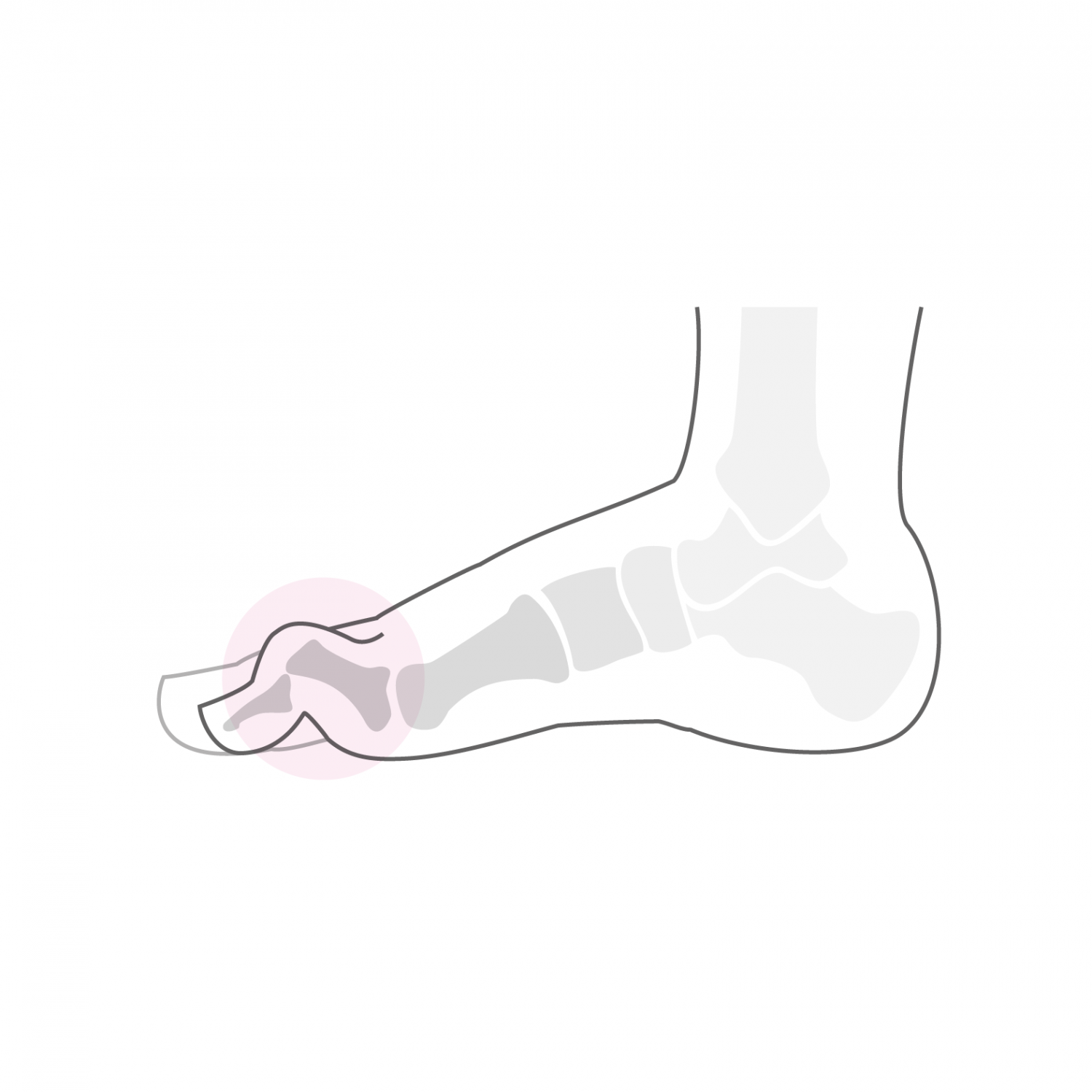
The position of the "little" toes depends on a balance between the extensor tendons which act on the top of the foot or dorsal side and the flexor tendons which are active on the bottom of the foot or plantar side. Their position is also dependent on the type of foot and a deformation most often localized on the 2nd toe is much more frequent in a "Greek" foot. In this type of foot, the 2nd toe is longer and therefore more exposed in the shoes and will tend to spontaneously develop flexion at the level of the first joint (proximal interphalangeal joint PPI). If the deformation is flexible at first and not disturbing, this position in prolonged flexion will cause a shortening of the articular capsule of the PIP joint and a contracture of the short flexor tendon which comes to be fixed at the base of the first phalanx. The deformation becomes more and more rigid, which will cause the development of an increasingly painful pressure point on the dorsal aspect of the toe, called the clavus. This flexure contracture of the PPI joint is accompanied over time by a contracture in extension of the metatarsophalangeal joint (joint at the base of the toe), we speak then of hammer toe. Under the continuous traction of the long flexor tendon of the toe which is fixed at the base of the 3rd phalanx develops a flexure contracture of the second joint (distal interphalangeal joint IPD) which results in the formation of a claw toe . The deformation of the toes is therefore progressive over time and passes from a "flexible" and straighten-able stage to a "rigid" and fixed position. It is often when the toe remains fixed in its deformation that the painful pressure points form. This hammer or claw deformation can affect an isolated toe or the 4 small toes simultaneously and it often also accompanies a hallux valgus situation, which makes the shoe more and more difficult. Other causes such as neurological or rheumatologic damage can also cause such deformities. In these cases all the toes are in principle concerned.
Treatment
The treatment is first of all conservative and consists of the adaptation of the shoes and the installation of plantar supports aiming above all to stabilize the situation. Care should be taken to wear shoes large enough to avoid pressure points. Daily exercises to mobilize the small joints and stretch the calf muscles also keep the toes moving for as long as possible.
If the pain and discomfort persist despite adequate conservative treatment, then surgical correction is required. This aims to correct all deformations in one time and aims to restore an adequate and harmonious alignment of the toes. We first perform a resection of the distal part of the 1st phalanx (Hohmann osteotomy), associated or not with a gesture on the extensor tendons aimed at correcting the dorsal extension of the articulation of the base of the toe . The corrected toe is fixed by a metal pin which will be removed at the consultation 3 weeks after the intervention. After this operation, full load walking in a shoe with a rigid sole is possible from the day after the operation and for a period of three weeks. If a hallux valgus is corrected at the same time, the shoe will be worn for 6 weeks.
After such an operation, the operated toes remain swollen for up to six months. From the 7th week, a physiotherapy treatment is set up in order to reduce this edema and restore mobility in the PIP and PID joints.
In case of associated metatarsalgia (diffuse pain under the heads of the metatarsal bones) which are often due to an excessive length of the 2nd, 3rd and 4th metatarsals, osteotomies of shortening of the metatarsals concerned are performed at the same time, in order to harmonize the support on the metatarsal heads. We take care to carry out these osteotomies in the intermediate 1/3 of the metatarsus (so-called diaphyseal osteotomy), in order to obtain a precise correction and an exact positioning of the metatarsal head and to avoid a stiffness of the articulation of the base of the toe, as is most often the case during sub-capital osteotomies (Weil osteotomies). We do not perform percutaneous osteotomies because this kind of osteotomy does not allow precise positioning of the metatarsal heads, which can cause an imbalance in the support of the forefoot and persistent pain.
In the case of diaphyseal osteotomies, the duration of wearing the shoe with rigid sole is 6 weeks. Once the osteotomies have been consolidated, walking is done gradually in a conventional shoe and a series of lymphatic drainage is prescribed to accelerate the recovery of the foot.
In these types of toe correction procedures, whether or not associated with other forefoot movements, the type of anesthesia and the length of stay in the clinic are discussed individually. All types of anesthesia are possible, from the simple peripheral block (anesthesia of the main nerve of the foot to the level of the hollow of the knee) to general anesthesia including spinal anesthesia. A pre-operative appointment is organized with our anesthesia service and the decision is made at that time. In case of correction of an isolated toe, the intervention can take place on an outpatient basis. However, you should know that this type of intervention can be quite painful in the immediate post-operative phase and that the treatment of this post-operative pain is more effective in a hospital setting. The foot needs rest in this immediate post-operative phase, this is to limit the development of hematoma (bleeding from operated tissues) and to promote healing of operative wounds. We therefore recommend carrying out the intervention on a short hospital stay with admission on the day of the intervention and a return home the next day or two days after the dressing has been repaired by the surgeon himself.